Yogesh Halekunche*1, Gopalakrishna Burdipad1, Sujatha Kuppusamy2, Suresh Janadri3
1Department of Pharmacology, R.R.College of Pharmacy, Bengaluru,Karnataka, India.
2Faculty of Pharmacy, Sri RamachandraUniversity, Porur, Chennai, Tamilunadu, India.
3 Department of Pharmacology, Acharya&BM Reddy College of Pharmacy, Bengaluru,
Karnataka, India.
*Address for Corresponding Author:
Mr. Yogesh H S
Assistant Professor
Department of Pharmacology
R. R. College of Pharmacy, Bengaluru,
Karnataka, India - 560 090
Tel: +91-9611133444
Abstract
Objective: To carry out the anti-osteoporotic activity of ethanolic extract of Punica granatum in ovariectomized rat model of osteoporosis at three different dose levels of 100, 300 and 500 mg/kg per day. Materials and methods: Healthy female albino rats were divided into six groups of six animals each. Group Iwas sham operated and served as control. All other groups were ovariectomized. Group II was fed with saline and served as ovariectomized control. Groups III–VI were orally treated with estrogen (2mg/kg) and ethanolic leaves extract of Punica granatum(100,300 and 500 mg/kg) respectively for 90 days. Results: The findings assessed on Punica granatum shows significant (P<0.001) increases in femur length, weight and density, significant (P<0.001) increases in serum calcium, phosphorus and reduction in alkaline phosphatase, tartrate resistant acid phosphatase, osteocalcin whereas urine calcium, creatinine and phosphorous levels were significant (P<0.001) decreasesand histology of femur exhibits restorative progress with increased ossification, mineralization and increased osteoclastic activity. Conclusion: Ethanolic extract of Punica granatum leaves shows anti-osteoporotic effect in OVX rats in dose depended manner.
Keywords: Osteoporosis, Punica granatum, Ovariectomy, Bone mineral density (BMD), Tartrate resistant acid phosphatase (TRAP), Osteocalcin (OC)
Introduction
Osteoporosis is a chronic, progressive disease of the skeleton characterized by bone fragility due to a reduction in bone mass and possibly to alteration in bone architecture that leads to susceptibility to fracture with minimum trauma (Kelly, 1996). The loss of bone has been attributed to an imbalance between bone formation and bone resorption. The type of osteoporosis associated with ovarian hormone deficiency following menopause is currently the most common cause of age associated boneloss (Arjmandi et al., 1996). Many synthetic agents including estrogens in hormone replacement therapy, selective estrogen receptor modulators like raloxifene, droloxifen, bisphosphonates and calcitonin, have been developed to treat osteoporosis but each of which associated with adverse events such as hypercalcemia, hypercalciurea, increased risk of endometrial and breast cancer, breast tenderness, menstruation, thromboembolic events, vaginal bleeding and hot flashes (Genant et al, 1998; Canalis et al.,1988; Gorman and Park, 2002). Hence, it would be most helpful to explore naturally occurring substances, especially of plant origin, that could prevent bone loss and are free from any adverse effects/events.
In most of the countries, incidence of osteoporosis about 2-4 times higher in women than the men. A sharp decrease in ovarian estrogen production is the predominant cause of rapid, hormone related bone loss during the first decade after menopause (Gruber et al., 1984). Menopause, aging and hereditary factors, inadequate calcium intake and absorption, lack of exercise, prolonged steroid administration, excessive alcohol intake, and cigarette smoking are the major risk factors that predispose osteoporosis (Prabha Shankar, 2002). The ovariectomized rat is a convenient and reliable model for studying the efficacy of pharmaceutical agents in post menopausal osteoporosis (Mitra et al., 2001; Bahram et al., 1996). The pharmacological agents used to manage osteoporosis act by decreasing the rate of bone resorption, thereby slowing the rate of bone loss, or by promoting bone formation. To overcome the wide range of side effects produced by these synthetic drugs, there is an increasing demand for green medicines which are thought to be healthier and safer for the treatment of osteoporosis. The phytoestrogens, which are known to bind to the estrogenic receptor sites of the cell and trigger the components and processes of estrogenic activities, have a promising role in the treatment of osteoporosis (Adams, 1989). The isoflavonoids are among the most active phytoestrogens in the flavonoid class. Ipriflavone, a synthetic flavonoid derivative (Agnusdei et al., 1989) was found to be effective in preserving bone mass in several models of experimental osteoporosis (Benvenutiet al., 1991). The isoflavones found in soybeans, such as genistein were found to prevent bone loss in the ovariectomized rat model of osteoporosis (Bahram et al., 1996; Blair, 1996; Fanti et al., 1998). Genistein was also found to suppress osteoclastic function by in vitro and in vivo (Blair, 1996).
P. granatum is one of the natural plant contain some phytoestrogens. Many years, Punica juice known and use for traditional medicine such as dried pericarp and the juice of the fruits are employed as orally medication in the treatment of colic, colitis, leucorrhea, menorrhagia, oxyuriasis, paralysis and external application to caked breast and to the nape of the neck in mumps and headache. Pomegranate juice is rich in antioxidants which general possess numerous important biological properties against cholesterol oxidation, atherogenesis, diabetes Alzheimer’s disease, act as anti-inflammatory and anti-aging (Sreekumar et al., 2012). However, the estrogenic effect of phytoestrogen as selective estrogen receptors modulator from P. granatum have not been investigated especially the tannin compound of pomegranates. Therefore, there is immense interest to investigate the effect and action of tannins of pomegranate on bone and reproductive tissue on osteoporosis model in rat.
Materials and methods
Collection and authentication of plant leaves
The medicinal plant P. granatum leaves collected from local market of Bengaluru, Karnataka during the period July 2012. The collected plant authenticated by Prof. P.V. Laxminarayana, Botanist, herbarium specimen kept in Pharmacology department of R. R. College of Pharmacy, Bangalore. The 5 kg of plant material dried properly, coarse powdered and stored in a well closed dark container.
Preparation of extract
Shade dried powdered of P. granatum leaves (5 kg) was exhaustively extracted with 95% ethanol using a soxhlet apparatus. The total ethanol extract was concentrated in vacuum to a syrupy consistency.
Experimental animals
Female Wistar albino rats weighing about 150–220 g in the age group of about 90 days were acclimatized to the experimental room at temperature 23±2°C, controlled humidity conditions (50–55%) and 12hour light/dark cycle. Animals were caged with a maximum of two animals each in a polypropylene cage and were fed with standard food pellets (Hindustan Lever Ltd., India) and water ad libitum. The study was conducted after obtaining ethical committee clearance from the Institutional Animal Ethics Committee of R.R. College of Pharmacy, Bangalore No. IAEC/RR/76/2012.
Acute toxicity
Female rats were fasted overnight before the test. The rats were received 5000 mg/kg of freshly prepared ethanolic extract of P. granatum leaves at single dose. The animals were observed immediately and then after 30 min, 1, 2, 4, 6 h and thereafter daily for 14 days. At the end of the fourteenth day the animals were sacrificed with excess ether as anesthesia and dissected for examination of vital organs (OECD guidelines 425 adoption).
Phytochemical screening
Phytochemical screening of the ethanol extract of leaves of P. granatum was carried out by standard procedures and tests (Trease and Evans, 1979) to find out the presence of chemical constituents such as alkaloids, terpenoids, flavonoids, tannins and coumarins.
Anti-osteoporotic activity
Experimental animals were divided randomly into six groups of six animals each. Group 1 was sham operated and served as control. All the other groups were ovariectomized and received treatment for 90 days starting from the fifteenth day of ovariectomized. Group II received vehicle and served as ovariectomized control. Group III was orally administered estrogen (2 mg/kg). Groups IV, V and VI were orally treated with suspension of ethanolic extract of P. granatum (ELPG) in 5% tween 80 at three different doses of 100, 300 and 500 mg/kg body weight respectively.
The rats were anesthetized with ketamine HCl (50 mg/kg, i.p.), and ovaries were removed bilaterally. Similarly sham operated animals were performed, but only exposing the ovaries. They were administrated with prophylactic gentamycin (10 mg/kg, i.p) for 3 days (Lasota and Danowask, 2004). The treatment of ethanol extract of P. granatum by oral gavages administration continued for 90 days. Body weight of all the rats was measured weekly (Reddy et al., 2004). At the end of 90 days, all the rats were deprived of food for overnight. On the next day, urine was collected and rats were anesthetized by ketamine HCl (50 mg/kg, i.p) and blood samples were withdrawn by retro-orbital plexuses. The blood samples were centrifuged at 2500rpm for 15 min to separate the serum and preserved (−20◦C) for analysis of serum calcium, phosphorus, alkaline phosphatase (ALP), tartarate resistant acid phosphatase (TRAP) and osteocalcin (OC) (Shirwaikar et al., 2003; Xie et al., 2005).Immediately, after collecting the urine and blood samples, uterus was carefully removed and weighed. The lumbar vertebra and femurs were isolated and stored at −70°C until biochemical, biomechanical and histopathological studies were performed.
Femur physical parameter
Fresh isolated left femurs were weighed using an electronic scale. Length of the femurs was measured using a digital slide calipers. The length was measured from the proximal tip of the femur head to the distal tip of the medial candyle (Reddy et al., 2004). Bone volume and density were measured by fluid displacement method (Asankursekhar et al., 2005).
Lumbar compression test
The fourth lumbar vertebra was identified and then isolated. The fresh vertebra was placed in digital hardness tester and compress until it got fractured, and the reading was recorded in Newtons (N) (Shirwaikar et al., 2003).
Femoral ash weight, ash percentage and ash calcium
After measuring the bone length of left femur it was placed in tared fused silica crucibles and kept in Muffles furnace (Growell Instrument, Bangalore, India) dried to a constant temperature at 600°C for 24 h. Then ash weight was determined and sample was suitably diluted with deionized water and assayed for calcium (Jiepin et al., 2009).
Biochemical estimation
The levels of serum calcium, phosphorous, ALP, TRAP and OC and urine calcium, inorganic phosphorus and creatinine were measured using semi-automatic analyzer with biochemical kits (Erba diagnostics, Mallaustr, Germany) (Shirwaikar et al., 2003; Xie et al.,2005).
Histopathology
The right femur was fixed in 10% formalin for 12 h at 4°C, decalcified in 5%EDTA for 7 days and embedded in paraffin wax and cut into sagittal plane section of5µmthickness of the femur. The sections were stained with hematoxylin and eosin (HE), and examined for histopathological changes under a light microscope (Bancroft and Cook, 1980).
Results
Effect of ELPG on body weight and uterine weight
There was no significant change in body weight in all the groups. The ovariectomy resulted in significant (P<0.001) decrease in uterine weight of OVX model group as compared to sham operated group (Table 1). Oral administration of 300 mg/kg and 500 mg/kg of ELPG shows significant(P<0.05) and (P<0.001) increase in uterine weight respectively, and estrogen group shows weight gain as compared to OVX model group and this difference was statically significant (P<0.001) after 90 days of treatment.
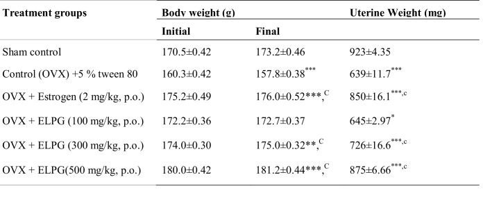
Table 1. Effect of ELPG on body weight and uterine weight in ovariectomized rats
All values are expressed as mean + SEM, n=6analyzed by One way Analysis of Variance (ANOVA) followed by multiple comparison Dunnet‘t’ test, aP<0.05, bP<0.01, cP<0.001 as comparison to Control (OVX) group, *P<0.05,**P<0.01,***P<0.001 as comparison to sham operated normal group.
Effect of ELPG on femur physical parameter
The length and weight of femur was decreased significant (P<0.001) in OVX model group as compared to sham operated group (Table 2). Following the administration of ELPG (100, 300 and 500 mg/kg) and estrogen in OVX rats significant increase in length, weight and density was observed which was comparable to the sham operated group. However, no significant changes in volume were observed in all the groups of animals. The dose dependent protection in the bone loss was observed in P. granatum leaves extract treated OVX rats.
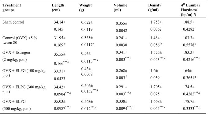
Table 2. Effect of ELPG on femoral length, weight, volume, density and 4th lumbar hardness in ovariectomized rats
All values are expressed as mean + SEM, n=6analyzed by One way Analysis of Variance(ANOVA) followed by multiple comparison Dunnet‘t’ test, aP<0.05, bP<0.01, cP<0.001 as comparison to Control (OVX) group, *P<0.05,**P<0.01,***P<0.001 as comparison to sham operated normal group.
Effect of ELPG on lumbar IV compression parameter
The lumbar vertebra hardness was decreased significant (P<0.001) in OVX group as compared to sham operated group (Table 2). After treatment with ELPG (100, 300 and500 mg/kg) and estrogen in OVX rats significant increases in biomechanical strength was observed which is comparable to sham group. The dose dependent protection was observed in P. granatum leaves extract treated OVX rats.
Effect of ELPG on biochemical parameters
The results of serum parameters in animals of different groupsare shown in (Table 3). The results indicates reduction in the serum calcium (P<0.001) and phosphorus (P<0.001) in OVX model group. In contrast, urine calcium, creatinine and phosphorous levels were significant elevated. P. granatum leaves extract (100, 300 and 500 mg/kg) and estrogen groups showed significant (P<0.001) dose dependent recovery of serum calcium and phosphorus, whereas urine calcium, creatinine and phosphorous levels were significant (P<0.001) decreases. Serum ALP levels elevated significant (P<0.001) in OVX model group as compared to sham operated group. In contrast, significant (P<0.001) reduction in levels of serum ALP levels were observed with ELPG (100, 300 and 500 mg/kg) and estrogen treated groups.
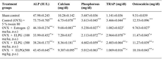
Table 3. Effect of ELPG on serum biochemical markers in ovariectomized rats
All values are expressed as mean + SEM, n=6analyzed by One way Analysis of Variance (ANOVA) followed by multiple comparison Dunnet‘t’ test, aP<0.05, bP<0.01, cP<0.001 as comparison to Control (OVX) group, *P<0.05,**P<0.01,***P<0.001 as comparison to sham operated normal group.
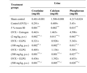
Table 4. Effect of ELPG on urinary markers in ovariectomized rats
All values are expressed as mean + SEM, n=6analyzed by One way Analysis of Variance (ANOVA) followed by multiple comparison Dunnet‘t’ test, aP<0.05, bP<0.01, cP<0.001 as comparison to Control (OVX) group, *P<0.05,**P<0.01,***P<0.001 as comparison to sham operated normal group.
Effect of ELPG on femoral ash weight, ash percentage and ash calcium
The femur ash weight, ash percentage and ash calcium were decreased significant (P<0.001) in OVX model group as compared to sham operated group (Table 5). In contrast, weight of ash, percentage of ash and calcium content was significant increase in ELPG (100, 300 and 500 mg/kg) and estrogen treated groups.
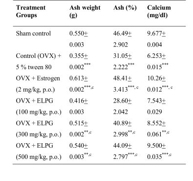
Table 5. Effect of ELPG on ash content of femoral bone in ovariectomized Rats
All values are expressed as mean + SEM, n=6analyzed by One way Analysis of Variance (ANOVA) followed by multiple comparison Dunnet‘t’ test, aP<0.05, bP<0.01, cP<0.001 as comparison to Control (OVX) group, *P<0.05,**P<0.01,***P<0.001 as comparison to sham operated normal group.
Histopathological evaluation
The femur section was examined for any histological changes. Epiphyseal region of Sham operated normal showing normal, compact and uniform trabecule with inter trabecular spaces (Figure 1A), Epiphyseal femoral region showing sparse, thinning of trabecule, loss of interconnectivity and widening of inter trabecular spaces, trabecule markedly disruptive, lytic changes and decrease in cells due to bone resorption (Figure1B), Epiphyseal femoral region showing moderately thick elongated trabecule and narrowed, last inter trabecular spaces and showing restoration of normal architecture and markedly diminished bone resorption by increasing bone cells (Figure 1C), Epiphyseal femoral region showing moderately, thick elongated trabecule and narrowed intertrabecular space and restoration of normal architecture along with increasing bone cells (Figure 1D), Epiphyseal femoral region showing moderately thick elongated trabecule, narrowed and compact Inter trabecular spaces associated with increased osteoblast cells (Figure 1E), Femoral region showing finely thick, elongated trabecule and narrowed, last Inter trabecular spaces showing complete restoration of normal architecture by forming cells (Figure 1F).Estrogen and ELPG (100, 300 and 500 mg/kg, p.o.) groups exhibited significant restorative progress with increased ossification, mineralization and increased osteoclastic activity and reduced bone resorption which indicates the recovery with essential features of normal bone.
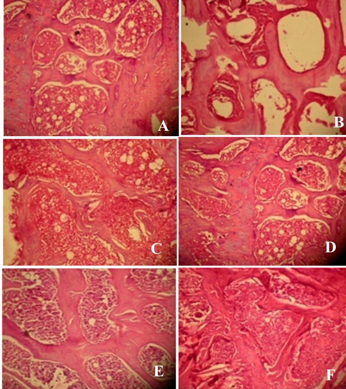 Figure1. Histopathological evaluation of ratsfemur sections. The sham group animals showed normal architecture and normal bone compactness (Figure 1A). The rats in OVX model group showed fragility with disruptive, lytic changes, thinning of the trabecule resulting in intertrabecular spaces widening (Figure1B). Estrogen and ELPG (100, 300 and 500 mg/kg) groups exhibited significant restorative progress with increased ossification, mineralization and increased osteoclastic activity and reduced bone resorption, which indicates the recovery with essential features of normal bone (Figure 1C–F).
Discussion
The aims of this work were to investigate the effects of the ethanolic extract of pomegranate leaves on bone protection and effects on reproductive organs. Ovariectomized rats are classically used as an animal model for studying the effect of postmenopausal bone loss. Furthermore, they may provide a useful model for investigating the biological effect of P. granatum on bone loss in ovariectomized rats. Pomegranate from ethanol of extract contain tannins especially ellagic acid (Sepulveda et al., 2011; Mori-Okamoto et al., 2004).
The pomegranate leaves comprising of various active compounds and these compounds were tannins as ellagic acid, gallotanic acid, alkaloids, glycosides, phenols, Saponins, coumarins, flavones and resins. Ellagic acid prevents bone loss by increasing mineralization of bone through osteoblast. The present study was investigated the potential preventive effects of tannin content ethanolic extract of P. granatum which contain ellagic acid to prevent bone loss in animal model of osteoporosis. The administration of ELPG prevented OVX-induced increase average body weight gain in rats. These results also support by previous study that compound that prevented OVX-induced uterine atrophy and increases in body weight gain, serum TRAP, OC, phosphorous and urine creatinine, calcium and phosphorous while significant decrease in ALP and calcium levels in serum. According with report, rats in the OVX group had lower densities of the right femur and tibiae because of reducing the ovariectomy induced increase in bone resorption. The administration of ELPG 100, 300 and 500 mg/kg effectively prevented OVX-induced lowering bone density. These observations are supported by previous study that ellagic acid significant prevented bone loss in OVX rats by increasing mineralization of bone (Bahtiar et al., 2014).
Osteoporosis, a metabolic bone disease results from a disturbance in normal bone remodeling, changing the balance to bone resorption over formation. Osteoporosis is caused by an imbalance between bone resorption and bone formation and results in bone loss and fractures after mineral flux. Estrogen-deficient OVX osteoporosis animal models have been used to evaluate osteoporotic drugs. Serum osteocalcin levels are generally considered to be a marker of bone turnover and serum ALP levels to be a marker of bone formation. As progression of OVX related osteoporosis, serum osteocalcin levels were generally increased along the increases of bone turn over, but serum ALP contents were decreased along inhibition of bone formations. On the contrary, the serum osteocalcin levels were decreased, but serum ALP levels were increased in present study, which suggested that the treatment activated the osteoblast differentiation and inhibited the bone mineralization and turn over. BMD provides information regarding the efficacy of anti-osteoporotic agents. Microscopic observations of bones also provide valuable information regarding bone morphology. In osteoporotic animals, the histological profiles are clearly changed compared with sham controls regardless of the cause, especially in the trabecular and cortical bones. The efficacy of various anti-osteoporotic agents has been evaluated using bone histology. Namely, some histomorphometrical indices for bone mass and bone formation are decreased concomitantly with increased bone resorption and this data can help predict the efficacy of anti-osteoporotic agents (Kang et al., 2015).
Many studies have shown that pomegranate juice and pomegranate polyphenol extracts can prevent many types of cancer, cardiovascular disease, diabetes, Alzheimer’s disease, arthritis, and colitis. Recent reports have shown that pomegranate seed oil and pomegranate juice contain several species of flavonoids and anthocyanidins, and that their antioxidant activity is three times more potent than that in red wine or green tea extract. The estrogenic activity of ELPG is mainly due to flavones and alkaloids, to a lesser extent, gallotanic acid. Flavones have various biological activities and can improve metabolic symptoms and bone protective effects in menopause. Flavonoids exert a weak estrogen-like effect by binding to ER-α and β in various tissues. Furthermore, flavonoids have the ability to interact with estrogen receptors and to control the activity of CYP19, an important enzyme in estrogen biosynthesis, and/or steroid dehydrogenases, (e.g., 11β-hydroxy steroid dehydrogenase). These effects induce various alterations causing a change in the overall hormonal balance, resulting in protection against bone loss and reducing osteoporotic effects and other menopausal symptoms. ELPG on postmenopausal symptoms are mainly due to the phytoestrogenic effects of flavonoids, gallotanic acid and alkaloids (Kang et al., 2015). Our present study shows that ELPG relieves climacteric symptoms through anti-osteoporotic activity. Further studies are needed to investigate the efficacy of that ellagic acid in humans.
Conclusions
The present studies suggest that a potential effect of ethanolic extract of P. granatum leaves increased the anti-osteoporotic effects in OVX rats on dose dependent manner. Therefore we suggest that pomegranate leaves are promising new potent protective agent for relieving osteoporosis in menopausal females.
Acknowledgments
The authors are thankful to principal and management of R. R. College of Pharmacy, Bengaluru for provided necessary facility to carry out the research work.
References
Adams NR. 1989. Phyto-oestrogens. In: Cheeke, P. (Ed.), Toxicants of Plant Origin. CRC Press, Boca Raton, FL, pp 23-51.
Agnusdei D, Zacchei F, Bigazzi S. 1989. Metabolic and clinical effects of ipriflavone in established postmenopausal osteoporosis. Drugs Experimental Clinical Research, 15(2): 97-104.
Arjmandi BH, Alekel L, Hollis BW, Amin D, Stacewicz SM, Guo P. 1996. Dietary soybean protein prevents bone loss in an ovariectomized rat model of osteoporosis. Journal of Nutrition, 126:161-167.
Asankursekhar D, Dolan D, Maitrayee M, Sandip M, Chandan M. 2005. Phytoestrogenic effect of black tea extract (Camellia sinensis) in an oophorectomized rat (Rattusnorvegicus) model of osteoporosis. Life sciences, 77:3049-3057.
Bahram HA, Lee A, Bruce WH. 1996. Dietary soybean prevents bone loss in a ovariectomized rat model of osteoporosis. Journal of Nutrition, 126:161-167.
Bahtiar A, Sunarmi A, Amalia R, Nurul Q, Puspita EW, Ade A. 2014. Polar Fraction of Punica granatum L. peel extract increased osteoblast number on ovariectomized rat bone. International Journal of Herbal Medicine, 2(1):65-70.
Bancroft JD, Cook HC. 1980. Manual of Histological Techniques. Churchill Livingstone, New York.
Benvenuti S, Tanini A, Masi L. 1991. Effects of ipriflavone and its metabolites on clonal osteoblastic cell line. Journal of Bone Mineral Research, 6:987-996.
Blair HC. 1996. Action of genistein and other tyrosine kinase inhibitor in preventing osteoporosis. Second International Symposium on the role of Soya in preventing and treating chronic disease. Brussels, Belgium 15-18 September.
Canalis H, McCarthy T, Centrella M. 1998. Growth factors and the regulation on bone remodeling. Journal of Clinical Investigation, 81:277-281.
Evans WC, Trease E. 1979. Pharmacognosy, 14th ed. Saunders, New Delhi.
Fanti P, Monier-Faugere MC, Geng Z, Schmidt J, Morris PE, Cohen D. 1998. The phytoestrogen genistein reduces bone loss in short term ovariectomized rats. Osteoporosis International, 8: 274-281.
Genant HK, Baylink DJ, Gallagher JC. 1989. Estrogens in the prevention of osteoporosis in post menopausal women. American Journal of Obstetrics and Gynecology, 161:1842-1846.
Gruber HE, Ivey JL, Baylink DL, Mathews M, Nelp WB, Sisom K, Chestnut CH. 1984. Long term calcitonin therapy in postmenopausal osteoporosis. Metabolism, 33: 295-303.
Jiepin W, Fujun S, Qibing M, Jianbo W, Rong Z, Siwang W. 2009. NO-donating genistein prodrug alleviates bone loss in ovariectomized rats. Swiss Medical Weekly, 138 (41-42): 602-607.
Kang SJ, Beom RC, Seung HK, Hae YY, Hye RP, Dong CK, Seong HC, Chang HH, Soo JP, Chang HS, Sae KK, Young JL. 2015. Dried Pomegranate Potentiates Anti-osteoporotic and Anti-obesity activities of red clover dry extracts in ovariectomized rats. Nutrients, 7: 2622-2647.
Kelly PJ. 1996. Is osteoporosis a genetically determined disease? British Journal of Obstetrics and Gynecology, 103: 20-27.
Lasota A, Danowask KD. 2004. Experimental osteoporosis different methods ovariectomy in female white rats. Roczniki Akademii Medycznejw Bialymstoku, 49: 129-131.
Mitra SK, Venkataranganna MV, VenkateshaUdupa U. 2001. The beneficial effect of OST-6 (OsteoCare), a herbomineral formulation in experimental osteoporosis. Phytomedicine, 8(3): 195–201.
Mori-Okamoto J, Otawara-Hama moto Y, Yamato H, Yoshimura H. 2004. Pomegranate extract improves a depressive state and bone properties in menopausal syndrome model ovariectomized mice. Journal of Ethnopharmacology, 92: 93-101.
Prabha Shankar B. 2002. Osteoporosis-a silent epidemic. Pharma Times, 34: 21-22
Reddy NP, Lakshmana M, Udupa UV. 2004. Anti-osteoporotic activity of OST-6, a herb mineral preparation in calcium deficient ovariectomized rats. Phytotherapy Research, 18: 25-29.
Sepulveda L, Ascacio A, Rodríguez-Herrera R, Aguilera-Carbo A, Aguilar CN. 2011. Ellagic acid Biological properties and biotechnological development for production processes. African Journal of Biotechnology, 10(22): 4518-4523.
Shirwaikar A, Khan S, Malini S. 2003. Anti-osteoporotic effect of ethanol extracts of Cissus quadrangularis Linn on ovariectomized rat. Journal of Ethnopharmacology, 89: 242-250.
Sreekumar S, Thankayyan R, Santhosh K, Baddireddi SL, Sreeharshan S. 2012. Pomegranate extract demonstrate a selective estrogen receptor modulator profile in human tumor cell lines and in vivo models of estrogen deprivation. Journal of Nutritional Biochemistry, 23: 725-732.


