Darshan Angadi1*, Omkarswamy Maradimath2, Ittagi Shanmukha2, Prashanth K. U.2
1Department of Pharmacology, SCS College of Pharmacy, Harapanahalli, Karnataka 583131 India
2Department of Pharamaceutical Chemistry, SCS College of Pharmacy, Harapanahalli, Karnataka 583131 India
*Address for Corresponding Author
Darshan Angadi
Department of Pharmacology
SCS College of Pharmacy, Harapanahalli, Karnataka 583131 India
Abstract
Objectives: The objective of study is to evaluate the Neuroprotective effects of stem bark extract of Calotropis gigantea Linn on experimentally induced neurotoxicity in rats. Material and methods: The various extracts were prepared using pet ether, 70% alcohol and water as solvents. After the completion of extraction, preliminary phytochemical study, quantification of antioxidant phytoconsutients like poly phenols, flavonoids and tannins were determined by spectroscopic method and further confirmed by HPLC method also. The extract rich in flavonoids, tannins and polyphenols (70% ethanolic extract) was selected for antioxidant and pharmacological studies. The 70% alcoholic extract of bark of Calotropis gigantea Linn (70% EEBCG) was subjected to pharmacological studies. The oxidative stress and inflammation has been well known in neurodegenerative disorders. Monosodium glutamate (MSG) and aluminium fluoride (AlF3) produces neuronal degeneration in animals. Oxidative stress mechanisms have been recently proposed in the MSG and AlF3 induced neurotoxicity. The compounds having antioxidant property have been suggested neuroprotection in different experimental models. Ethanolic extract of Calotropis gegantea linn bark (EEBCG) is known for its potent antioxidant activity; hence the present study has been designed to evaluate the neuroprotective effect of EEBCG on MSG and AlF3 models. Results: In the present study, 70% EEBCG showed protection against MSG and AlF3 induced neurotoxicity. The antioxidant property of EEBCG may be responsible for its neuroprotective action.
Keywords: Ficus religiosa Linn; antioxidant: Monosodium glutamate, Aluminium fluoride, excitotoxicity, oxidative stress, neurodegeneration
Introduction
Neurodegenerative sicknesses arise whilst nerve cells in the mind or peripheral nervous gadget lose function over the years and in the long run die. Although remedies may help relieve some of the bodily or mental symptoms related to neurodegenerative sicknesses, there's currently no way to sluggish sickness development and no recognized cures (Markesbery, 2007).
Common neurodegerative issues incorporate of dementias like Alzheimers ailment [AD]. Parkinsons sickness, Huntingtons disease and cerebrovascular Stroke. Increased oxidative pressure is the essential underlying purpose for neurovascular harm. Other contributing elements for these disorders encompass diabetes mellitus, hypertension, hyperlipidemia, more suitable platelet aggregation and coagulation hobby, ischaemic heart disorder, atherosclerosis, endothelial dysfunction and hyperhomocysteinemia (Scarmeas et al., 2006).
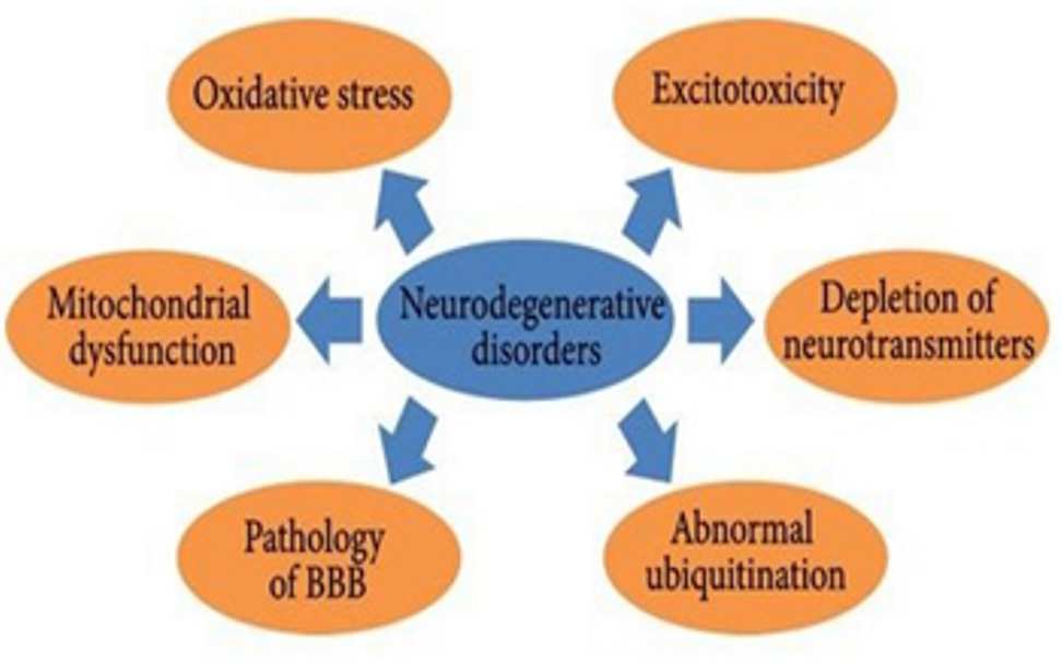
Figure 1. Various causes of neurodegenerative disorder
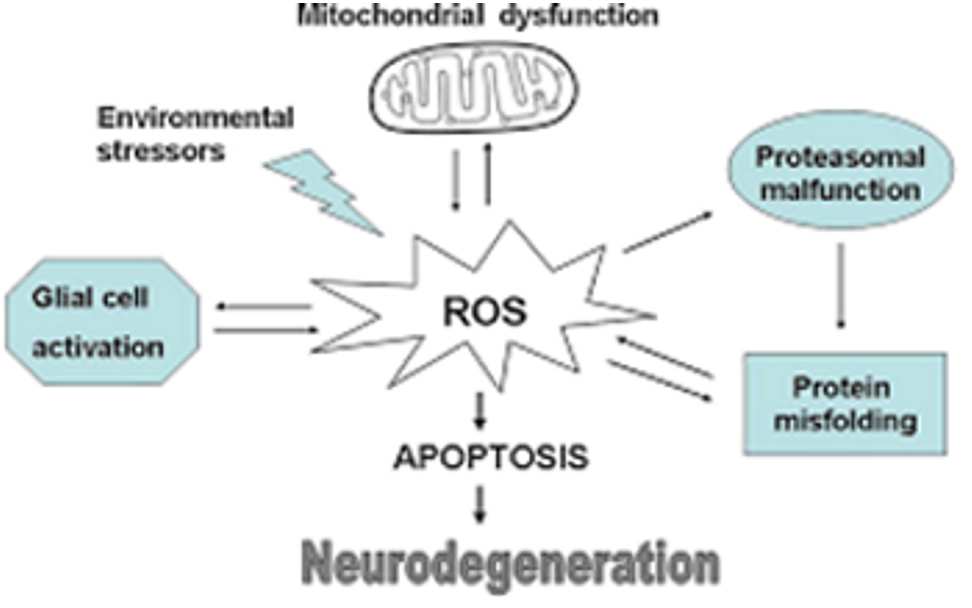
Figure 2. Pathogenesis of neurodegenerative disorder
Antioxidants are materials which can prevent or gradual damage to cells due to free radicals, volatile molecules that the frame produces as a reaction to environmental and other pressures. They are sometimes referred to as "unfastened-radical scavengers." The assets of antioxidants can be natural or artificial.
Calotropis gigantea Linn is a tall shrub reaching 2.4-3m high; returned yellowish white, furrowed; branches stout, terete, more or much less covered (specially the more youthful ones) with great apprised cottony (Dai et al., 2006), pubescence. Leaves 1-20 with the aid of 3.eight-10 cm. Flowers inodourous, purplish or white. Calyx divided to the base; sepals 6 by way of four mm, ovate, acute, cottony (Claiborne, 1985). It contain numerous phytochemical constituents along with Giganteol, α and β calotropeol, β-amyrin, Calotropnaphthalene, calotropisesquiterpenol, calotropisesterterpenol (terpene derivatives), Sapogenins, holarrhetine; cyanidin-3-rhamnoglucoside; taraxasterol, isovalerate (Gasior et al., 2006).
Calotropis gigantea Linn belongs to the family Asclepiadaceae, commonly known as Safed aak, Aak, Alarkh, Madar, Sveta Arka and Akanda. Its roots contain cardiac glycosides, seven oxypregnane-oligo glycosides, calotroposides A-G. Root bark contains β- amyrin, two isomeric crystalline alcohols, giganteol, isogiganteol and cardenolides. Latex reported to contains akundarin, latex contains 0.45% uscharin, 0.15% calotoxin, 0.15% calactin, latex also contains α- calatropeol, β- calotropeol, β- amyrin and calcium oxalate, it also yields a nitrogen and sulphur containing fish and cardiac poison, gigantin. Latex also contins traces of glutathione and a proteoclstic enzyme similar to papain. Leaves are reported positive for alkaloids, glycosides, mudarine. Stembark: β- calotropeol, β-amyrin, giganteol. Flowers are consists of α- calatropeol, β-calotropeol, amyrin, cardioactive glycosides, mudarine, asclepin, bitter resins akundarin and calotropin. Stem Bark of plant contains Giganteol, α and β calotropeol, and β-amyrin.
Calotropis gigantea traditionally used as asthma, abortifacient, analgesic and antinociceptive activity, antifertility and emmenagogue, anti-inflammatory activity, anthelmintic activity, anti-cancer activity, anti dote for scorpion stings and insect bites, anti tumor activity, anti-diarrheal and anti dyssentry activities, antimicrobial activity, antiviral activity, anxiety and pain, cns activity, cytotoxic activity, dyspepsia, elephantiasis, epilepsy, elephantiasis of the legs and scrotum.
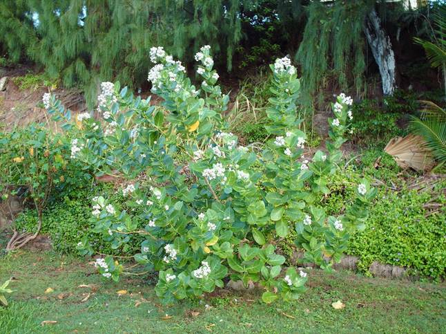
Figure 3. The leaves of Calotropis gegantea
Material and methods
Plant Material
Bark of calotropis gegantea Linn was collected from surroundings of Harapanahalli, Karnataka. The plant was identified and authenticated by Prof. K. Prabhu, Department of Pharmacognosy, S.C.S. College of Pharmacy, Harapanahalli.
Animals
Wister albino rats (weighing 150-250g) of either sex were used in this study. They were procured from Sri Venkateshwara Enterprises, Bengaluru. The animals were acclimatized for one week under laboratory conditions. They were housed in polypropylene cages and maintained at 27°C ± 2°C under 12 hrs dark / light cycle. They were fed with standard rat feed (Gold Mohur Lipton India Ltd.) and water ad labium was provided.
Pharmacological activities
Mono sodium glutamate induced Neuro toxicity
The animals were divided into five groups of six rats each as follows and treated for 7 days as follows;
Group 1: Received normal saline (i.p.) + vehicle (p.o.).
Group 2: Received MSG 2 g/kg (i.p.) + normal saline (p.o.).
Group 3: Received MSG 2g/kg (i.p.) + ascorbic acid 100 mg/kg, (i.p.),
(reference standard).
Group 4: Received MSG 2 g/kg (i.p.) + 70% EEFRB 250mg/kg (p.o)
Group 5: Received MSG 2 g/kg (i.p.) + 70% EEFRB 500mg/kg (p.o)
The gap between 70% alcoholic extract and MSG is 1 hour. The dose of MSG was selected based on previous literature. During the drug treatment, rats were observed for the behavioral changes for 50 minutes daily (Rang et al., 2003). On 8th day the rats were evaluated for body weight change, locomotor activity, balance beam task, Elevated plus maze and rota rod test. On 9th day rats were sacrificed and brains were isolated for estimation of glutathione, SOD, CAT, lipid peroxidation and total protein (TP).
B) Aluminum fluoride induced Cerebrotoxicity
The animals were divided into five groups of six rats each as follows;
Group 1: Received normal saline (i.p.) + vehicle (p.o.).
Group 2: Received (AlF3−) 600ppm + normal saline (p.o.).
Group 3: Received (AlF3−) 600ppm + ascorbic acid 100 mg/kg, (i.p.)
Group 4: Received (AlF3−) 600 ppm + 70% EEFRB 250mg/kg (p.o).
Group 5: Received (AlF3−) 600 ppm + 70% EEFRB 500mg/kg (p.o).
Animals from each group were fed with drug or vehicle for 10 days prior to aluminum fluoride treatment through their drinking water (7 days at a dose of 600 ppm). During the drug treatment, rats were observed for the behavioral changes for 50 minutes daily (Kulkarni et al., 2021). Rats were evaluated for body weight change, locomotor activity, balance beam task, Elevated plus maze and rotarod test. Later rats were sacrificed and brains were isolated for estimation of glutathione, SOD, CAT, lipid peroxidation and total protein (TP).
Behavioral parameters
The spontaneous locomotor activity was monitored using actophotometer (Dolphin Pvt. Ltd., Mumbai, India) equipped with infrared sensitive photocells, the apparatus was placed in darkened, light and sound attenuated and ventilated testing room. Before locomotor task, animals were placed individually in the activity meter for 2 min for habituation (Claiborne, 1985). Thereafter, locomotor activity was recorded for a period of 5 min. The locomotor activity was expressed in terms of total photo beam counts per 5min.
B) Rota rod test
The Rota rod test was used to measure the muscle relaxant property of the rats. Turn on the roto-rod. Appropriate speed 20-25rpm selected, rats were placed one by one on the rotating rod (Tiwari, 2001). Fall of time was noted, the cut of time of Rota rod test is 5min.
C) Beam walking task for motor coordination
The Beam walking task for motor coordination was used to measure the ability of rats to traverse a horizontal narrow beam (2.3 cm × 120 cm) suspended 50 cm above a foam-padded cushion (Tietz, 1970). During testing, the rats were given 1 min to traverse the beam. If they did not complete the task or if they fell off the beam, the trial was ended and the rats were placed back into their home cages. For successful performers, the latency to cross the beam was recorded.
Biochemical estimations
A) GSH estimation
Tissue samples were homogenized in ice cold trichloroacetic acid (0.5G tissue plus 5 ml, 10% TCA) in an ultra turrax tissue homogenizer. Briefly, after centrifugation at 3000 rpm for 10 minutes, 0.5 ml supernatant is added to 2 ml of 0.3 M disodium hydrogen phosphate solution. A 0.2 ml solution of dithiobisnitrobenzoate (0.4 mg/ml in 1% sodium citrate) is added and the absorbance at 412 nm is measured immediately after mixing. Percent increase in OD is directly proportional to the increase in the levels of glutathione. Hence, % increase in OD was calculated.
B) In vivo lipid peroxidation
Stock solution of TCA-TBA-HCl reagent: 15% w/v trichloroacetic acid; 0.375% w/v thiobarbituric acid; 0.25N hydrochloric acid was prepared. Take 1.0 Stock solution of TCA-TBA-HCl reagent: 15% w/v trichloroacetic acid; 0.375% w/v thiobarbituric acid; 0.25N hydrochloric acid was prepared. 2.0 ml of TCA-TBA-HCl was added and mixed thoroughly. The solution was heated for 1 hr in a boiling water bath. After cooling, the flocculent precipitate was removed by centrifugation at 1000 rpm for 2 min. The absorbance of the sample was determined at 535 nm against a blank.
C) Catalase (CAT)
Catalase measurement was done based on its ability to decompose hydrogen peroxide (H2O2). Briefly, to 1.95 ml of 10 mM H2O2 in 60 mM phosphate buffer (pH = 7.0), 50 µl of the brain tissue supernatant was added and the rate of degradation of H2O2 was followed at 240 nm per min. % increase in OD is directly proportional to the increase in the levels of Catalase (James et al., 2005). Hence, % increase in OD was calculated.
D) Super oxide dismutase (SOD)
Superoxide dismutase activity was determined based on the ability of SOD to inhibit the auto-oxidation of epinephrine to adrenochrome at alkaline pH (Misra and Fridovich, 1972). Briefly, 0.5 ml of the supernatant obtained from the centrifuged brain homogenate was added to a mixture of 0.1mM epinephrine in carbonate buffer (pH 10.2) in a total volume of 1 ml and the formation of adrenochrome was measured at 295 nm (James et al., 2005). % increase in OD is directly proportional to the increase in the levels of Super Oxide Dismutase. Hence, % increase in OD was calculated.
E) Total protein
The total serum protein contents were determined by Biuret’s method (using total protein kit). This method is based on the principle that proteins give an intensive violet-blue complex with copper salts in an alkaline medium. Iodine is included as an anti-oxidant. The intensity of the colour formed is proportional to the total protein concentration in the sample.
Results and Discussion
Plants are conceived as source of antioxidants due to presence of poly phenols, tannins and flavonoids, which possess wide spectra of biological properties. Recent studies showed that many tannin, flavonoids and related polyphenols contribute significantly to the total antioxidant activity. Antioxidants further helps as organ protectants. Hence, in this study a widely grown calotropis gegantea Linn reported to possess polyphenolic compounds. The preliminary phytochemical screening of plant showed that, they possess polyphenols, flavonoids, tannins and vitamins which are reported to possess antioxidant property. These antioxidant properties may be assessed by several in-vitro models like Super oxide radical and reducing power assay and was selected for screening. The barks were collected and extracts has prepared separately and were subjected to preliminary phytochemical studies, the results indicated that bark possess tannins, flavonoids, polyphenols and vitamins in 70% ethanolic and aqueous extracts. Since 70% ethanolic and aqueous extracts contain similar type of phytoconstituents, those two extracts were subjected to the quantification. The total phenol, flavonoid, tannin content of 70% Ethanolic was 55.20mcg/ml, 152.33mcg/ml and 2538.88mcg/ml expressed as equivalent to catechol, quercetin and tannic acid respectively, which is better compared to aqueous extract. Ethanolic extract of bark showed dose dependent super oxide anion scavenging and reducing power activity. Therefore 70% ethanolic extract of bark of calotropis gegantea was selected to screen neuroprotective activity. Results of the present study indicates that calotropis gegantea bark extract treatment significantly improved motor deficits, muscle grip strength, body coordination and restored the body weight, hind limb functions and antioxidant levels in brain.
Table 1. Total phenolic, flavonoid and tannin content of extract
|
Particulars |
Phenolic content |
Flavanoid content |
Tannin content |
|
Standard curve |
catechol |
Quercetin |
Tannic acid |
|
Web length (nm) |
650nm |
510nm |
700nm |
|
Amount of content in 70 percent alcoholic content |
55.20mcg /ml |
152.33mcg/ml |
2538.88mcg/ ml |
|
Amount of content in aqueous content |
45.31mcg /ml |
38.33mcg/ml |
638.88mcg/ml |
|
R2 value |
0.998 |
0.980 |
0.998 |
Table 2. 70% EEBCG on body weight change and behavioural change in MSG treated rats
|
Treatment |
% Body weight change |
Rota Rod Test (s) |
Locomotor Activity (Count/5 min) |
Balance beam test (sec) |
|
Negative control |
- |
3955±190.2*** |
280.8±18.59*** |
28.84±4.777*** |
|
Positive control |
13.04 |
1454±250.9 |
92.17±19.19 |
13.45± 3.232 |
|
Standard (Ascorbic acid) |
04.80 |
3936±204.7*** |
274.3±19.19*** |
28.25±3.232*** |
|
250mg/kg 70%EEBCG |
12.18 |
2931±221.4** |
204.2±17.37** |
25.33± 3.064* |
|
500mg/kg 70%EEBCG |
08.24 |
1712±151.7NS |
137.0 ±44.39NS |
18.26± 1.533NS |
Each value is expressed as mean ± SEM (n = 6), where, NS represents non significant; ****P<0.0001 – highly significant;***P<0.001 – very significant; **P<0.01- good significant; *P<0.05- significant, when compared to MSG alone treated rats. One-way ANOVA followed by Dunnett’s comparison test.
Table 3. Effect of 70% EEBCG on biochemical parameters in MSG treated rats
|
Treatment |
GSH |
LPO |
SOD |
CAT |
TP |
|||||
|
Mean± SEM |
%increase |
Mean± SEM |
%increase |
Mean± SEM |
%increase |
Mean± SEM |
%increase |
Mean± SEM |
%increase |
|
|
Negative control |
0.148±0.001*** |
- |
0.125± 0.001 **** |
- |
0.282± 0.009 **** |
- |
0.832± 0.001 **** |
-
|
0.653± 0.006 **** |
----- |
|
Positive control |
0.076±0.001****
|
- |
0.491± 0.001 **** |
----- |
0.111± 0.009 **** |
----- |
0.363± 0.001 **** |
- |
0.372± 0.006 **** |
----- |
|
Standard (Ascorbic acid) |
0.137±0.001***
|
80.26 |
0.158± 0.005 **** |
67.82 |
0.241± 0.005 *** |
90.47 |
0.712± 0.003 **** |
96.14 |
0.599± 0.011 **** |
61.0 |
|
250mg/kg 70% EEBCG |
0.106±0.001 ****
|
39.47 |
0.188± 0.002 **** |
61.71 |
0.027± 0.004 ****
|
86.48 |
0.672± 0.0008 **** |
85.12 |
0.541± 0.008 ****
|
45.43 |
|
500mg/kg 70% EBCG |
0.077±0.002 *** |
1.31 |
0.219± 0.002 **** |
55.39 |
0.178± 0.002 **** |
60.36 |
0.555± 0.001 **** |
52.89 |
0.490± 0.008 *** |
31.72 |
Each value is expressed as mean ± SEM (n = 6), where, NS represents non significant; ****P<0.0001 – highly significant;***P<0.001 – very significant; **P<0.01- good significant; *P<0.05- significant, when compared to MSG alone treated rats. One-way ANOVA followed by Dunnett’s comparison test.
Histopathological Studies in MSG induced neurotoxicity
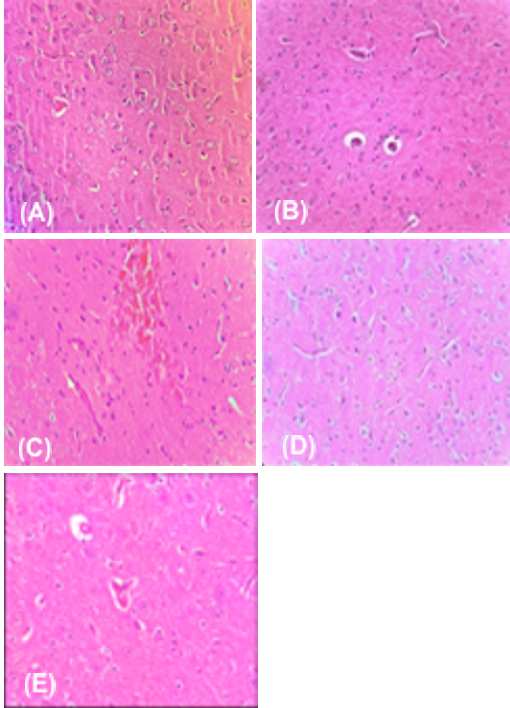
Figure 4. Photomicrograph of brain parenchyma tissue from MSG induced neurotoxicity: (A) Negative control (B) Positive control (C) Standard (D) Lower dose (250 mg/kg) of 70% EEBCG (E) Higher dose (500 mg/kg) of 70% EEBCG
Table 4. Effect of 70% EEBCG on body weight change and behavioural change in AlF3 treated Rats
|
Treatment |
%body weight change |
Rotarod test(s) |
Balance beam test(s) |
Locomotor activity (count/5min) |
|
Negative control |
----- |
1764±58.36**** |
28.84±3.716* |
459.3±25.41*** |
|
Positive control |
11.04 |
846.2±36.09 |
11.98±1.072 |
212.33±27.06 |
|
Standard (Ascorbic acid) |
05.80 |
1625±36.62**** |
26.81±3.491** |
432.3±33.83** |
|
250mg/kg 70% EEBCG |
12.18 |
1554±34.39**** |
25.27±1.968 |
350.8±24.29** |
|
500mg/kg 70% EEBCG |
8.24 |
1062±85.08NS |
17.28±1.415* |
263.7±27.99NS |
Each value is expressed as mean ± SEM (n = 6), where, NS represents non-significant; ****P<0.0001 – highly significant;***P<0.001 – very significant; **P<0.01- good significant; *P<0.05- significant, when compared to MSG alone treated rats. One-way ANOVA followed by Dunnett’s comparison test.
Table 5. Effect of 70% EEBCG on biochemical parameters in AlF3 treated rats
|
Treatment
|
GSH |
LPO |
SOD |
CAT |
TP |
|||||
|
Mean± SEM
|
%increase
|
Mean± SEM |
%increase |
Mean± SEM |
%increase |
Mean± SEM |
%increase |
Mean± SEM |
%increase
|
|
|
Negative control |
0.1485± 0.0012**** |
----
|
0.1252± 0.0014 **** |
----- |
0.2827± 0.0096 **** |
----- |
0.8327± 0.0064 **** |
-----
|
0.6535± 0.0047 **** |
----- |
|
Positive control
|
0.0525± 0.0011**** |
----
|
0.4917± 0.0004 **** |
----- |
0.01117± 0.0042 **** |
----- |
0.4302± 0.0061 **** |
----- |
0.3807± 0.0033 **** |
----- |
|
Standard (Ascorbic acid) |
0.1373± 0.0010**** |
80.65
|
0.1588± 0.0045 **** |
67.7 |
0.2413± 0.0035 **** |
96.0 |
0.7065± 0.0088 **** |
64.22 |
0.5912± 0.0073 **** |
55.29 |
|
250mg/Kg 70% EEBCG
|
0.1255± 0.0016****
|
65.13 |
0.1775± 0.0124 **** |
63.9 |
0.2113± 0.0054 *** |
84.42 |
0.6502± 0.0070 **** |
51.13 |
0.5372± 0.0091 |
41.1 |
|
500mg/Kg 70% EEBCG
|
0.1070± 0.0031**** |
40.78
|
0.2198± 0.0038 **** |
55.29 |
0.1878± 0.0060 *** |
68.12 |
0.6503± 0.0071 **** |
40.7 |
0.4778± 0.0081 *** |
25.5 |
Histopathological Studies in ALF3 induced neurotoxicity
D) 250mg/kg 70%EEBCG: Treatment with 70% 250mg/kg EEBCG in AlF3 showed the section studied from the brain parenchyma shows intact architecture. Few of the pyramidal cells and neuroglial cells show degenerative changes with moderate inflammatory infiltration (Figure 5D). Most of the blood vessels show moderate perivascular inflammatory infiltration.
E) 500mg/kg 70%EEBCG: Treatment with 500mg/kg of EEBCG in AlF3 induced rats section studied from the brain parenchyma shows intact architecture. The pyramidal cells and neuroglial cells appear intact with moderate inflammatory infiltration (Figure 5E). Few of the blood vessels show mild perivascular inflammatory infiltration.
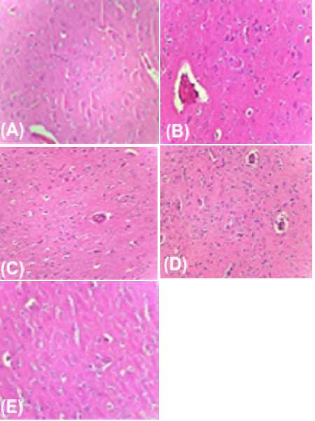
Figure 5. Photomicrograph of brain parenchyma tissue from in ALF3 induced neurotoxicity: (A) Negative control (B) Positive control (C) Standard (D) Lower dose (250 mg/kg) of 70% EEBCG (E) Higher dose (500 mg/kg) of EEBCG
In present the study sub-chronic administration of MSG (2 g/kg, i.p. for 7 days) and AlF3 (600ppm p.o for 7 days) produced significant decrease in body weight, motor and muscle grip related behaviors and antioxidant status in the brain. These findings are well co-relates to histopathological observations which indicates the presence of degenerated nerve cell and loosening of nerve fibers in striatal region of the brain. The reduction of body weight in MSG and AlF3 induced rats could be due to metabolic impairment caused by neurotoxic agents i.e. impairment in energy metabolism, mobilization of energy stores and lipid peroxidation which constitute peripheral effects.
Striatal lesion indicated by treatment with MSG and AlF3 caused an impairment in the locomotion observed in actophotometer. MSG and AlF3 have been reported to cause lesion in hippocampal and pyramidal neurons. Animals showed poor locomotion in the treated groups. This observation indicates that the treatment with MSG and AlF3 causes motor dysfunction as observed in the PD patients. In the present study EEFRB treatment significantly improved locomotor performance and showed improved motor activity. Since GSH is considered as inbuilt antioxidant substance which prevents lipid peroxidation, estimation of tissue GSH and extent of lipid peroxidation were considered as parameters of screening in-vivo antioxidant properties in all the models. Since the MSG induced neurotoxicity was reported to be via free radicals, the neuroprotective activity of test extract in this model is also attributed to the antioxidant activity of the plant. The neuroprotective property of the extract is further confirmed by significant improvement of the brain architecture by reversing the disintegrated neuropil fibres over MSG group. The neuroprotective property of the extract is further confirmed by significant improvement of the brain architecture by reversing the disintegrated neuropil fibers over AlF3 group. In the present study, it was observed that the bark possess polyphenolic compounds (flavonoids and tannins) and these constituents are reported to have antioxidant and organ protective properties. Hence the anti-oxidant and neuroprotective properties may be attributed to the polyphenolic constituents that are present in the bark of Calotropis gegantea Linn.
From the results and discussion, it will be concluded that, the plant possesses significant quantity of phenols, flavonoids and tannin with very good antioxidant properties. Phenolic components are known to be antioxidants and antioxidants are reported to have organ protective role. Conclusively, the 70% ethanolic extract of calotropis gegantea Linn bark has neuroprotective potential against monosodium glutamate and aluminium fluoride induced neurotoxicity in rats. EEFRB attenuates the behavioral impairment and oxidative stress induced by MSG and AlF3. It also prevented the neurodegeneration in striatal and hippocampal regions due to MSG and AlF3 toxicities. The observed protective effects may be due to antioxidant properties of EEFRB. However, further research is required to elucidate the specific mode of action.
References
Claiborne A. Handbook of methods for oxygen radical research. London CRC Press. 1985: 283–4.
Dai Q, Borenstein AR, Wu Y, et al. 2006. Fruit and vegetable juices and Alzheimer’s disease: the Kame Project. Am J Med 119:751–759.
Gasior M, Michael AR, Adam LH. 2006. Neuroprotective and Disease-modifying Effects of the Ketogenic Diet. Behavioural Pharmacology 17(5-6): 431-39.
James WS, Wang J, Wang X, Evelyn P, Laszlo P, James AD. 2005. Mitochondria Play a Central Role in Estrogen-Induced Neuroprotection. CNS & Neurological Disorders - Drug Targets 4:69-83.
Kulkarni SK. Hand book of experimental Pharmacology. 3rd edition Nirali publisher, Pune 2021.
Markesbery W. 2007. Damage to Lipids, Proteins, DNA, and RNA in Mild Cognitive Impairment. Archives of neurology, 64(7):954-956.
Ramanathan M, Sivakumar S, Anandvijaykumar PR, Saravanababu C, Rathinavel Pandian P. 2007. Neuroprotective evaluation of standardized extract of Centella asciatica in monosodium glutamate treated rats. Indian J Experimental Biology, 45: 425-431.
Rang HP, Dale MM, Ritter JM, Moore PK. Pharmacology. 5th ed. Edinburgh: Churchil Livingstone. 2003: 490-3.
Rekha R, Saleem BN, Ruby S. 2009. Evaluation of in-vitro antioxidantactivityof stem-bark and stem wood of premna serratifolia. Research Journal of Pharmacognosy and Phytochemistry, 1(1): 11- 14.
Scarmeas N, Stern Y, Tang MX, et al. 2006. Mediterranean diet and risk for Alzheimer’s disease. Annals of Neurology, 59:912–921.
Sharma SS, Gupta S. 2007. Neuroprotective effect of MnTMPyP, a superoxide dismutase/catalase mimetic in global cerebral ischemia is mediated through reduction of oxidative stress and DNA fragmentation. European J Pharmacology. 561: 72-9.
Tietz NW. Fundamentals of clinical chemistry. W.B. Saunders Co Philadelphia PA. 1970: 302
Tiwari KA. 2001. Imbalance in antioxidant defence and human disease. Current Sciennce 81: 1179-86.


